

.png)

.png)







Home
 News Center
3D Scanning Imaging of Brain Neural Networks: High-Precision Motion Stages Enhancing Accuracy and Efficiency
News Center
3D Scanning Imaging of Brain Neural Networks: High-Precision Motion Stages Enhancing Accuracy and Efficiency
News Center
3D Scanning Imaging of Brain Neural Networks: High-Precision Motion Stages Enhancing Accuracy and Efficiency
3D Scanning Imaging of Brain Neural Networks: High-Precision Motion Stages Enhancing Accuracy and Efficiency
 2026-06-03
Share:


"Coretech's ART series high-speed, high-precision motion stages are widely used in 3D scanning imaging equipment for biological tissues, particularly in brain neural network scanning imaging applications, thanks to their outstanding speed stability and minimum step capability."
01 Brain Neural Network Research
The brain is the pinnacle of biological evolution, and deciphering its working mechanisms is one of humanity's ultimate aspirations. To date, however, scientists have yet to reveal the fundamental mechanisms underlying brain functions such as memory, thought, and consciousness. The limited understanding of brain structure and function also hampers the development of effective drugs and therapies for brain disorders such as Alzheimer's disease and Parkinson's disease.
The mammalian brain is a highly complex network consisting of millions to billions of densely interconnected neurons. The cell bodies of neurons, arterioles, and venules are only several tens of micrometres in diameter, capillaries are only a few micrometres in diameter, and dendrites and axonal fibres are 1 μm or less in diameter.

In previous studies, researchers classified brain neurons into different types based on cellular morphology, electrophysiological characteristics, neuronal projections, and combinations of expressed molecules and transcriptomic profiles. These different types of neurons are distributed across distinct cortical layers and perform diverse functions.
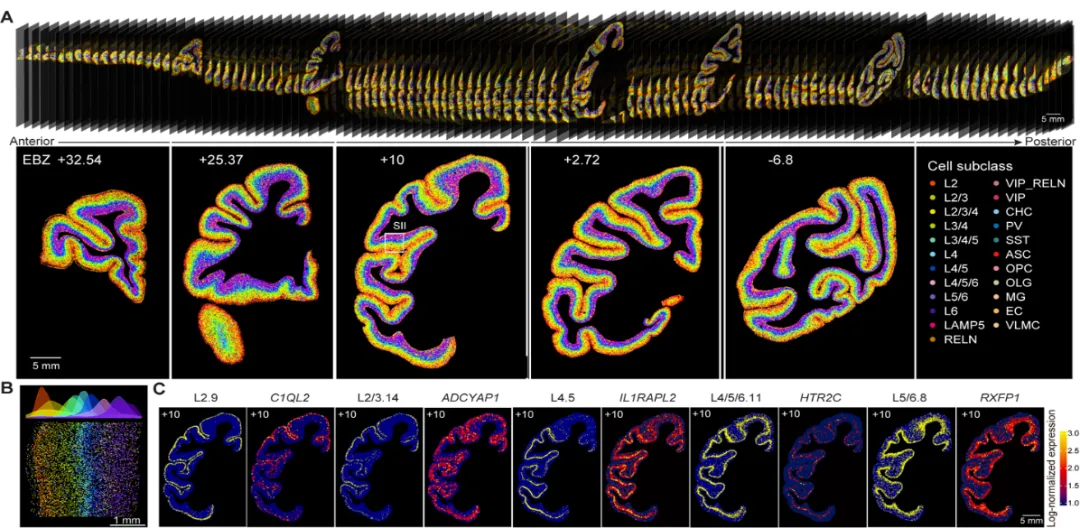
A more direct and effective research approach is to map the three-dimensional fine structures of neural circuits and vascular networks across the entire brain at the mesoscopic scale. This would provide critical structural information for understanding the brain and is a crucial prerequisite for elucidating the operational mechanisms of brain functions. Producing a complete brain cell distribution atlas would propel neuroscience research to an entirely new level, and has thus become a key battleground for neuroscientists worldwide.
02 3D Neural Network Imaging Technology
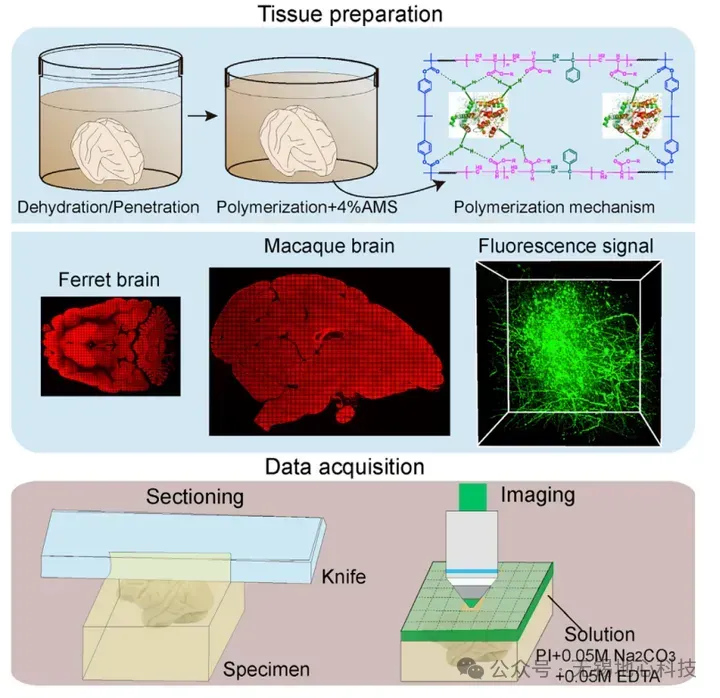
The Micro-Optical Sectioning Tomography (MOST) system, invented by Professor Luo Qingming's team at Huazhong University of Science and Technology, is a whole-brain imaging system based on mechanical sectioning. This system uses an imaging system to image the sample surface, while a microtome removes the surface tissue of the plastic-embedded sample. By continuously imaging new surfaces, the system ultimately acquires a complete mouse brain dataset with axial resolution of 1 μm.
For fluorescently labelled samples, Professor Luo's team developed a series of fluorescence MOST (fMOST) systems, including single-photon fMOST, two-photon fMOST, and the brain-wide positioning system (BPS) based on structured illumination. Among these, the single-photon fMOST and two-photon fMOST systems employ point-scanning imaging, which requires long imaging times. The BPS system uses structured illumination for faster imaging, with sectioning and imaging performed separately; it uses a mercury lamp as the light source, enabling rapid, high-resolution whole-brain imaging. This system is capable of not only imaging at the neural circuit level but also acquiring fine neuronal morphology.
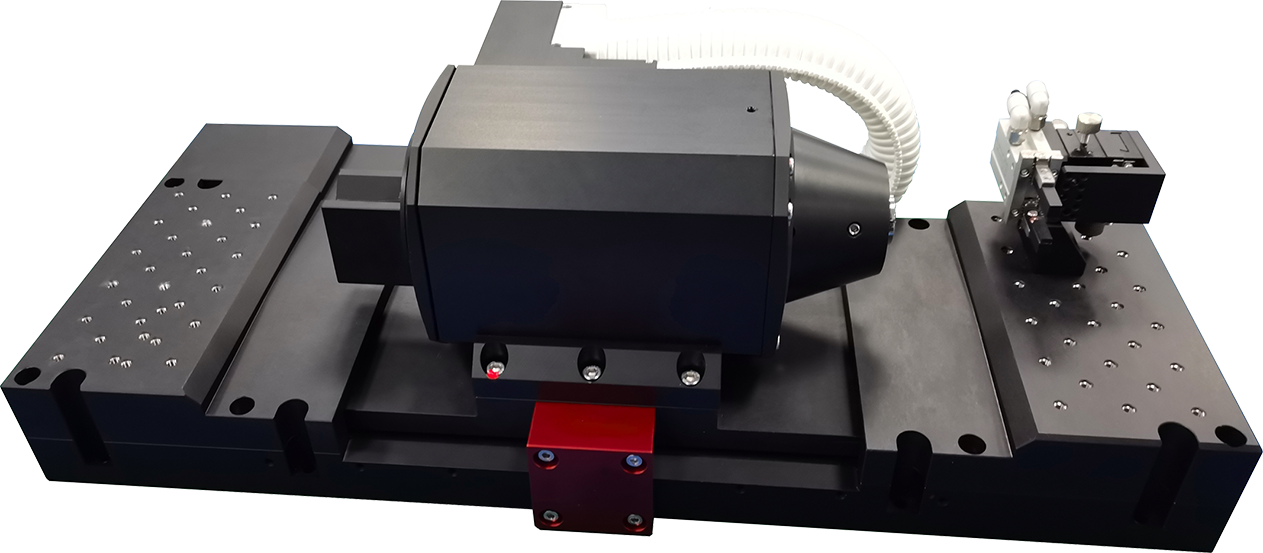
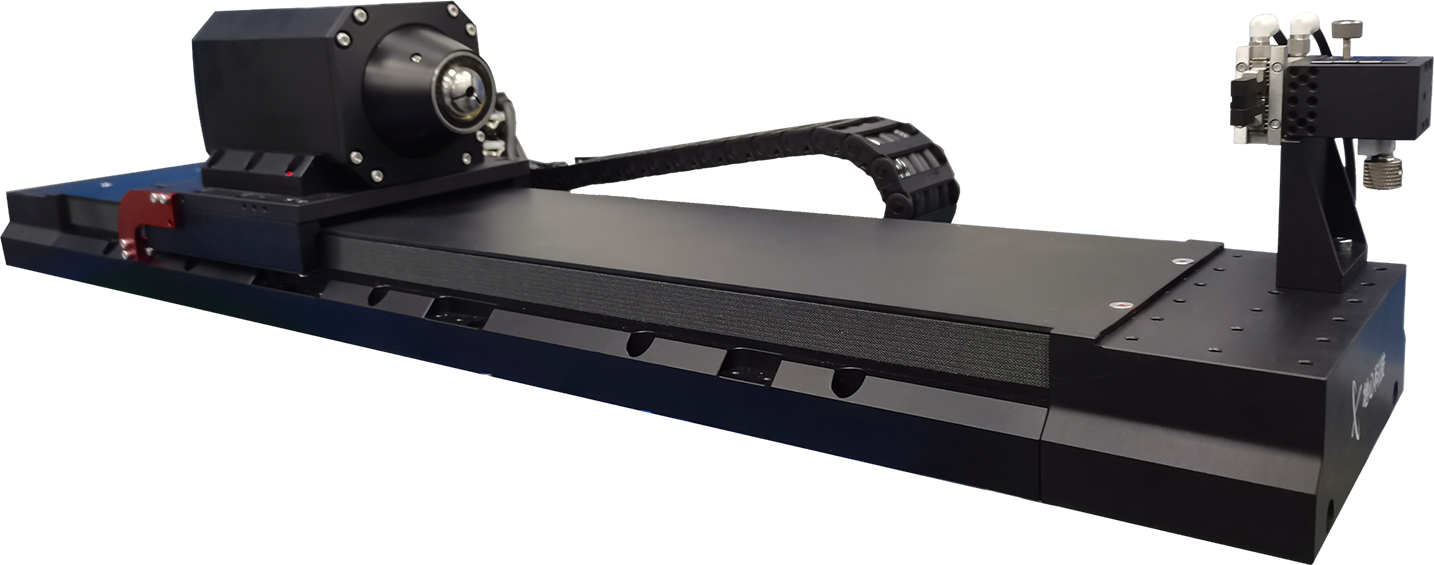
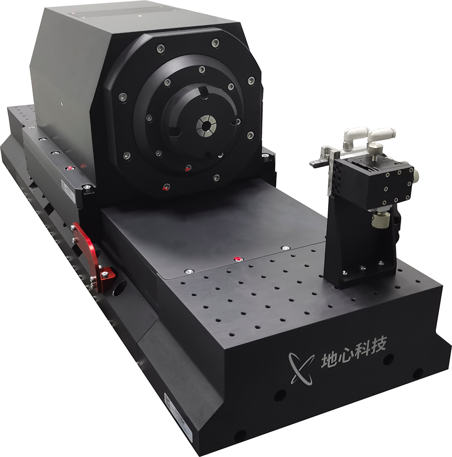
Taking neuronal process imaging in whole-brain imaging as an example, to obtain high-resolution structural information of long-range or local neural circuits over large volumes or the entire brain, it is necessary to establish a structured-illumination fluorescence microscopy imaging system with sub-micrometre resolution. This imaging technique can capture fine structures such as long-range axons and dendrites while simultaneously providing spatial localisation information of cellular architecture. Additionally, this imaging system can acquire vascular network information across the whole brain. A typical whole-brain imaging BPS system is illustrated below.
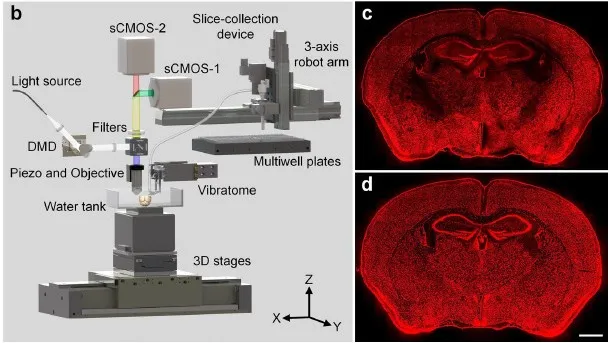
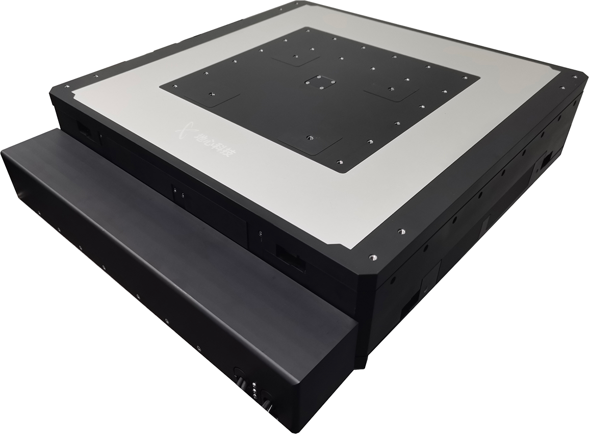
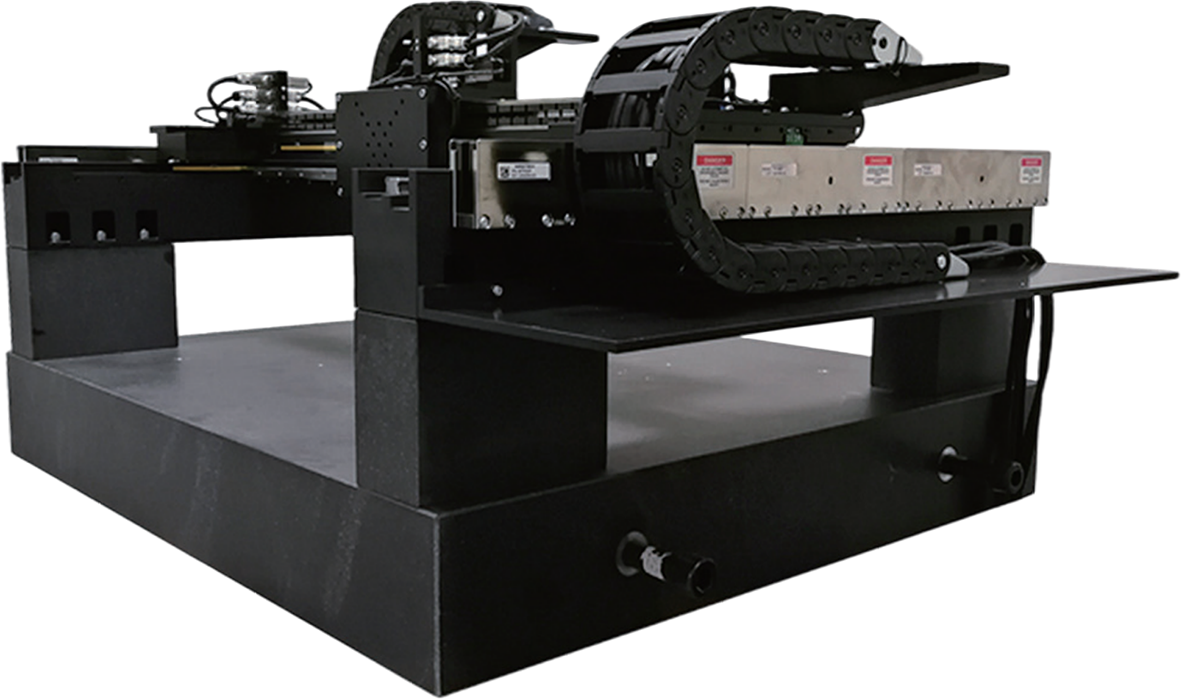
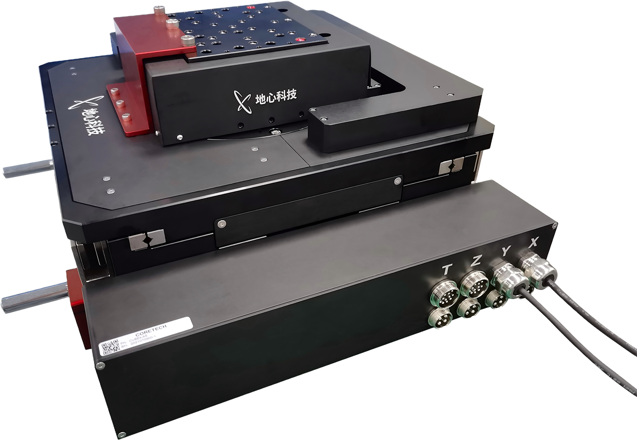
High-precision 3-axis precision drive control technology is one of the key technological aspects of whole-brain imaging equipment. Acquiring and tracing long-range neural circuits across the entire brain at micrometre resolution presents a major challenge. Coretech's nano-positioning stages and high-load, high-stiffness Z-lift stages perfectly meet the various demanding requirements of such applications, including:
Sub-micrometre motion step increments
Nanometre-level in-position stability
Stage shear stiffness for lateral blade sectioning
Long-term trouble-free operation over days or even weeks (MTBF)
Synchronised triggering of motion and image acquisition
03 Coretech Products
Coretech has many years of application experience in the medical equipment industry, particularly in neural network scanning imaging technology. A variety of product combinations can meet the needs of different users. Over many years of deployment, the company has not only accumulated extensive application experience but has also earned a strong reputation.
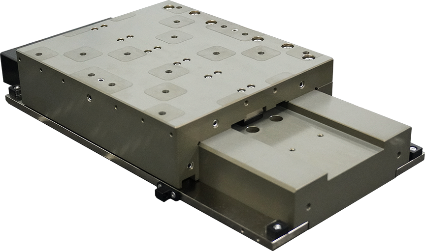
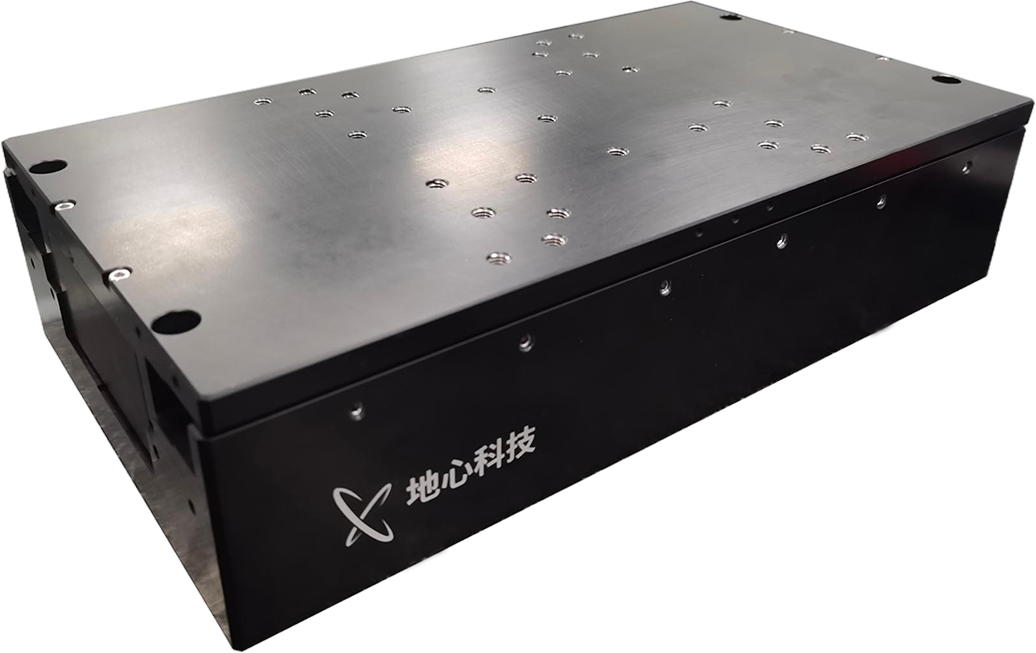
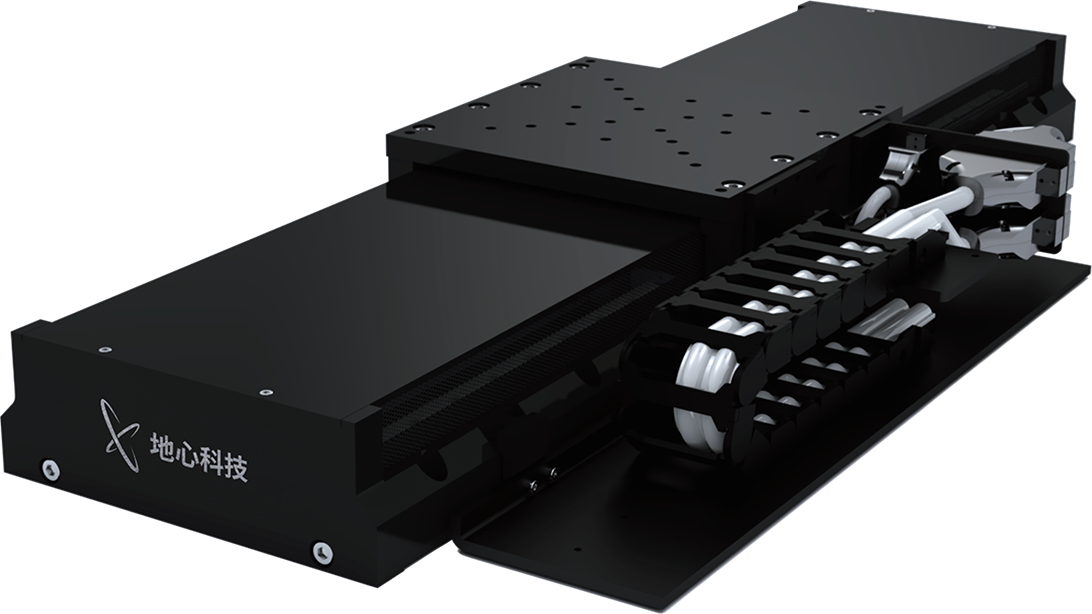
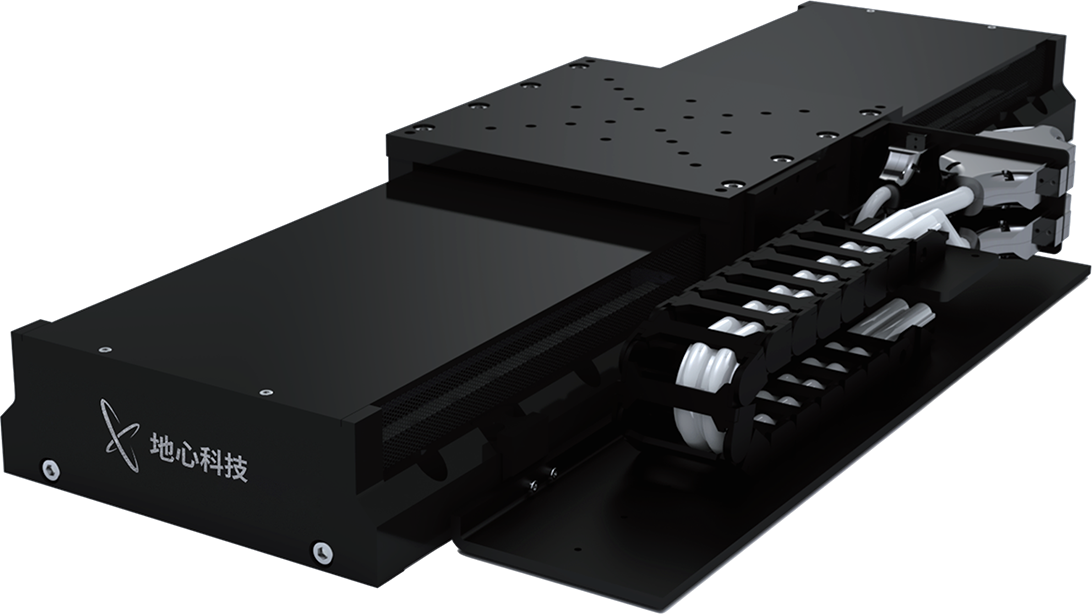
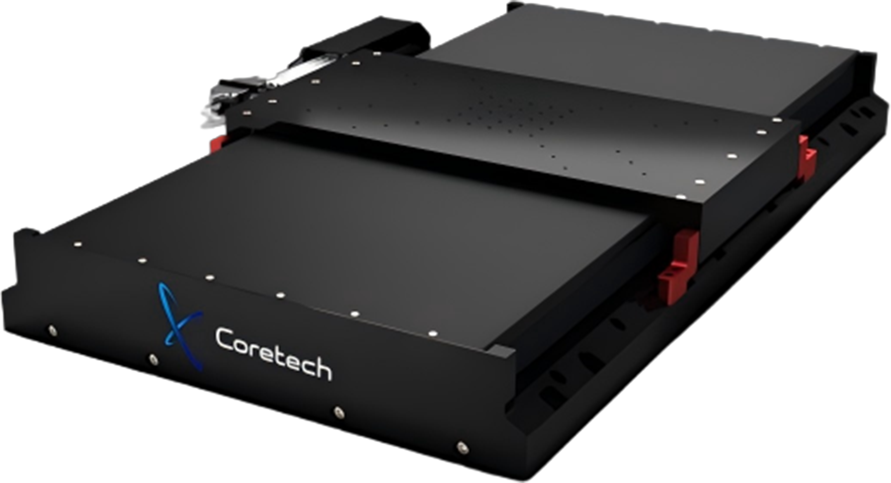
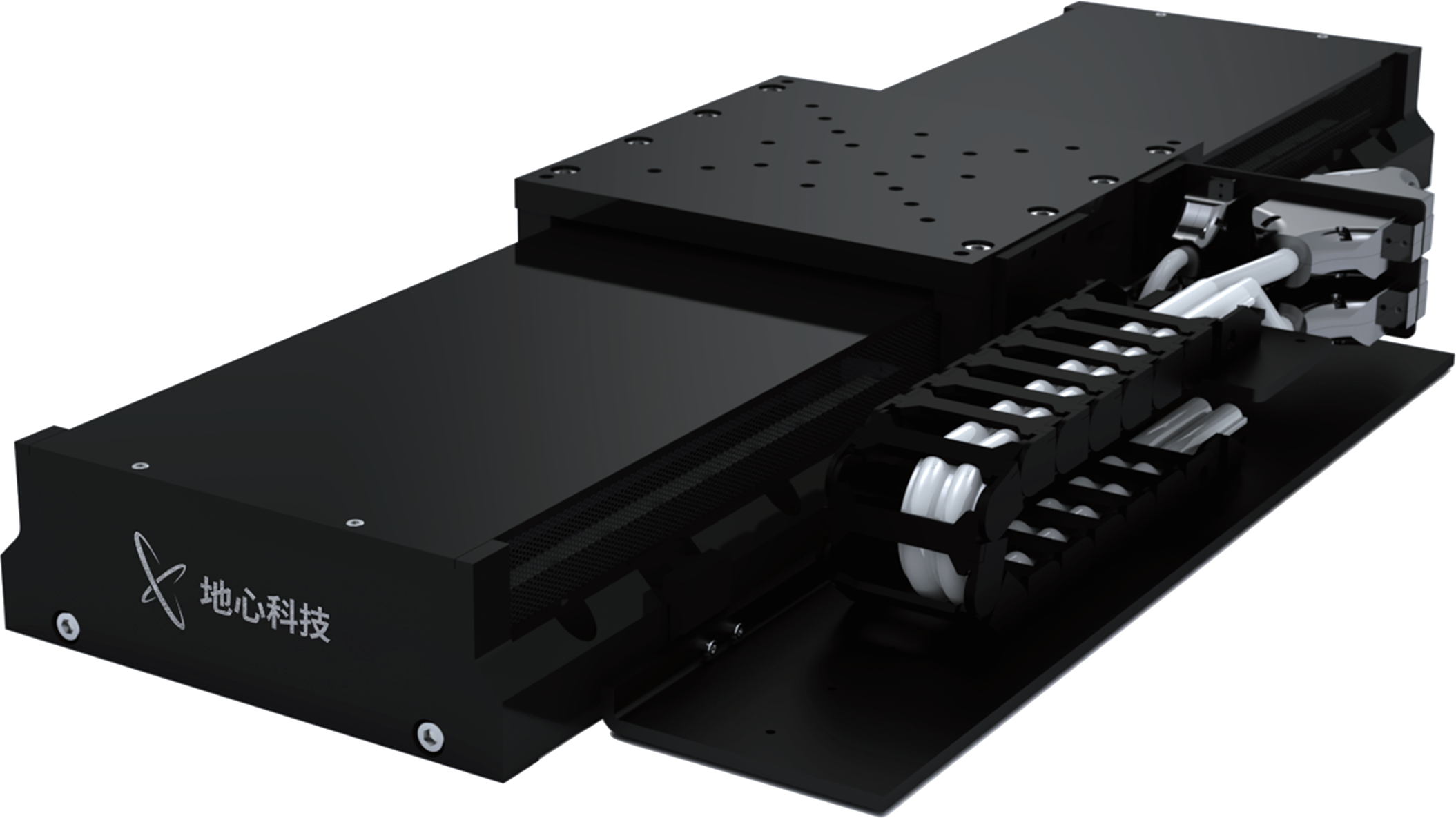
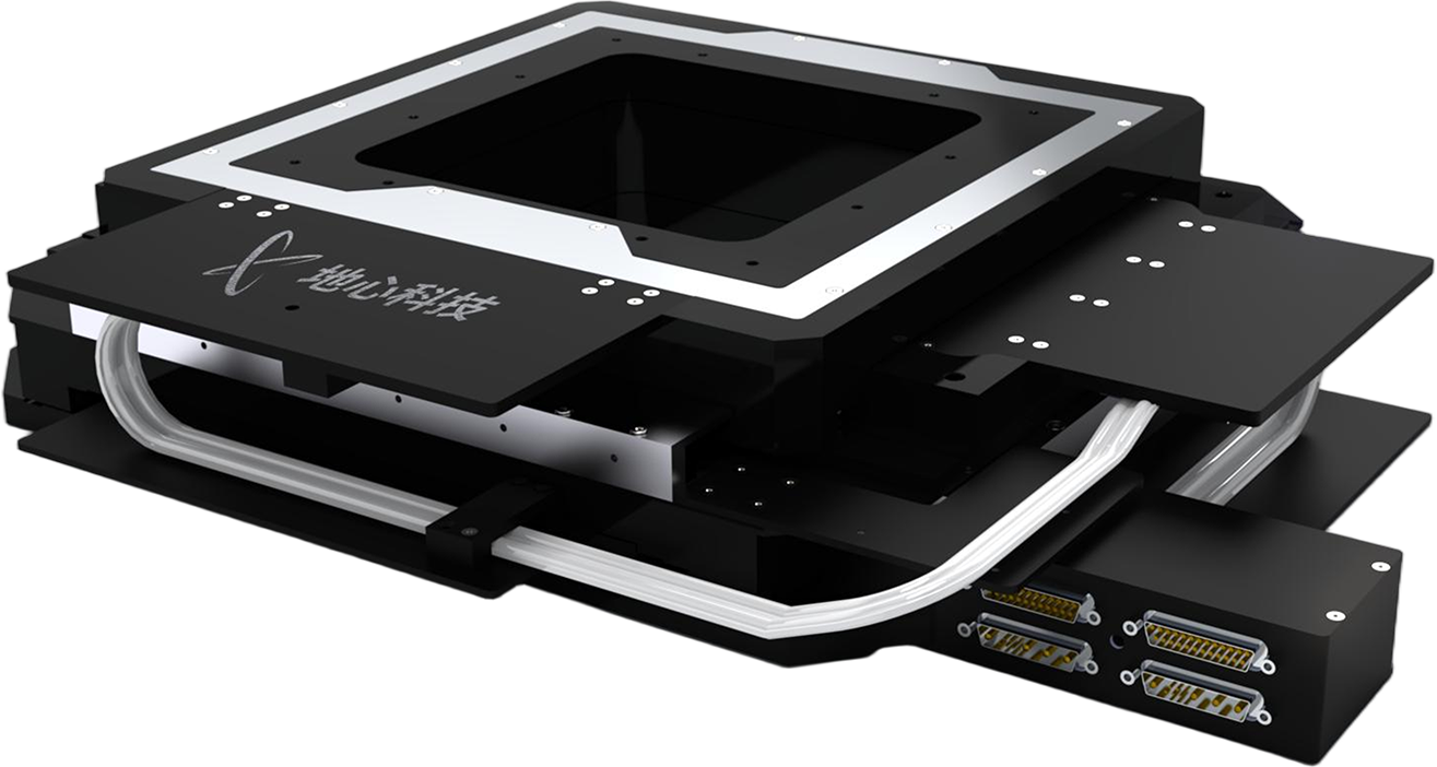
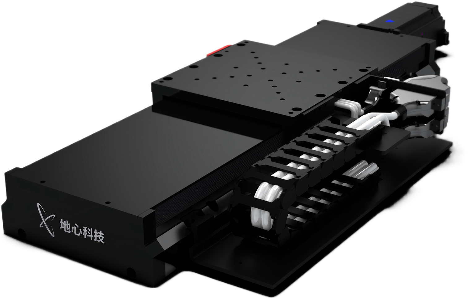
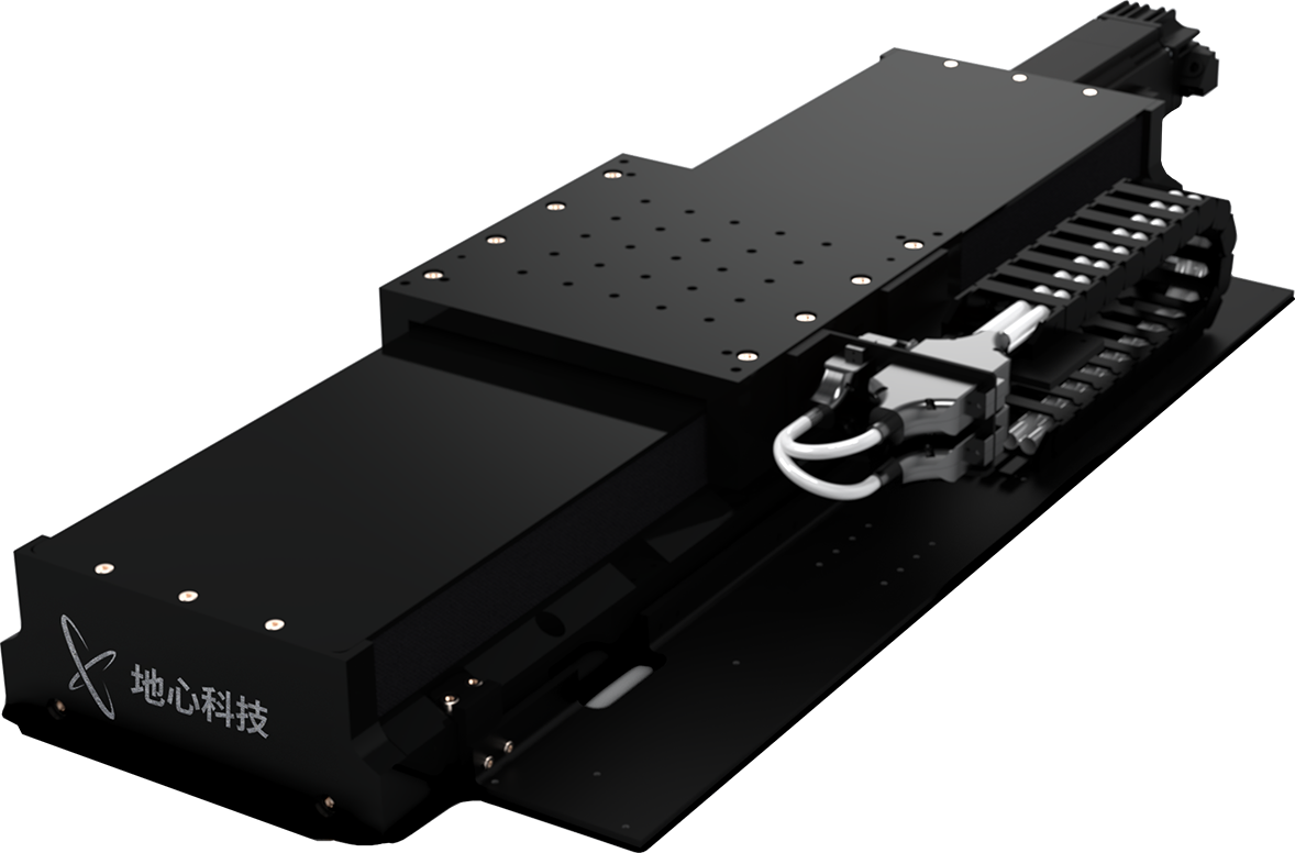
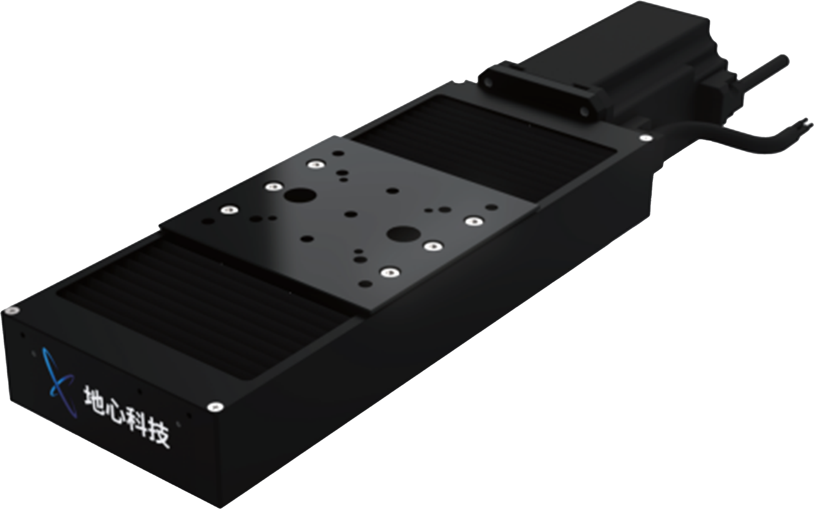
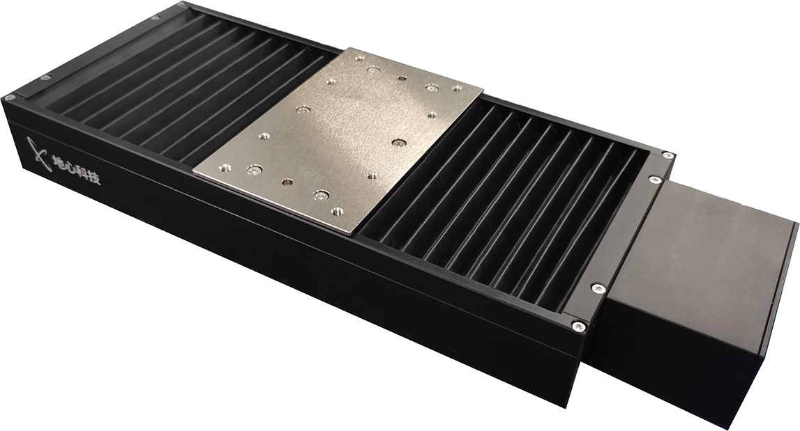
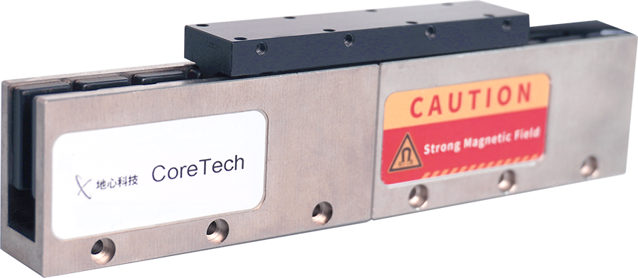
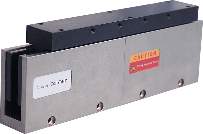
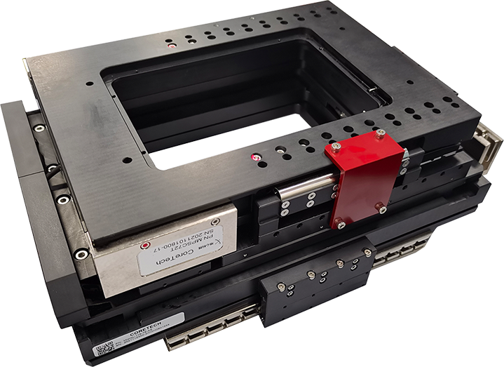
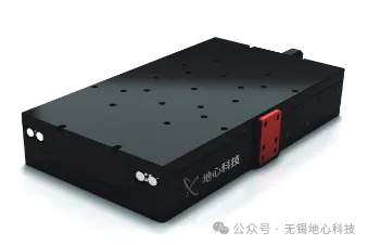
ART130 Series Linear Stage
As a small-step stepping axis, the cross-roller bearing stage offers excellent motion and settling times.
High-density position-triggered camera capture at 5 nm intervals ensures high-resolution, distortion-free images.
Flexible travel configuration options provide abundant product choices for different sample sizes.
Nanometre-level in-position stability enables sub-micrometre probe density precise positioning and printed layer resolution.
Stage cutoff frequency >100 Hz enables fast motion and settling.
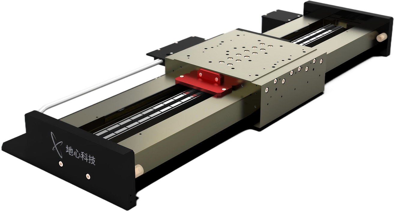
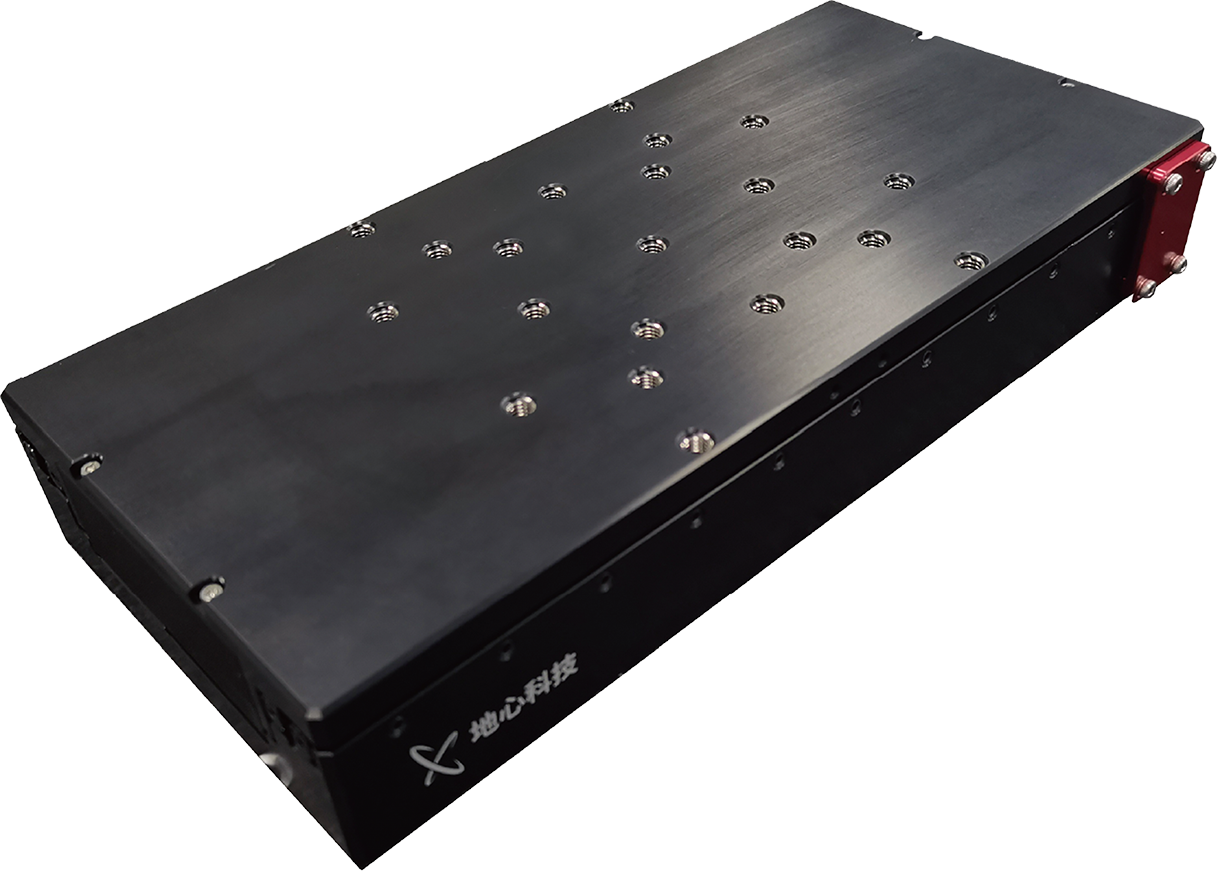
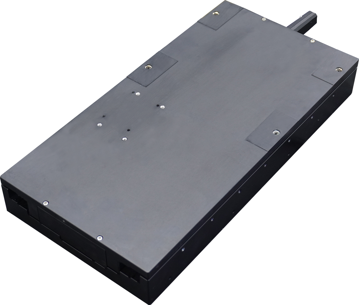
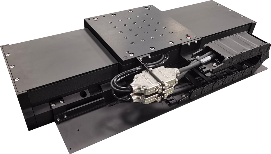
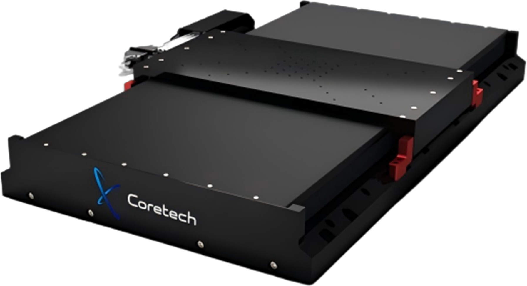
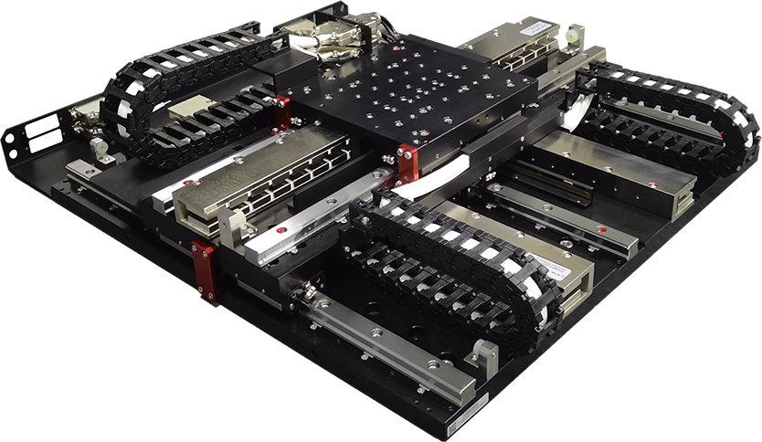
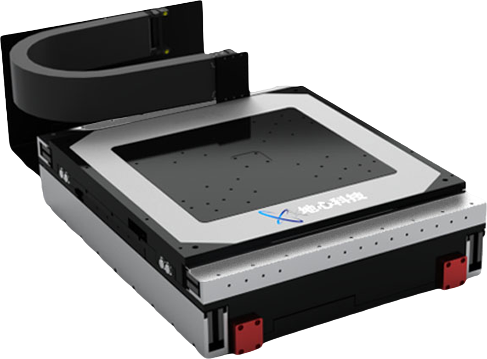
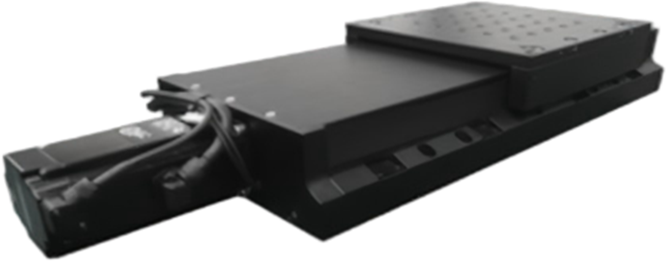
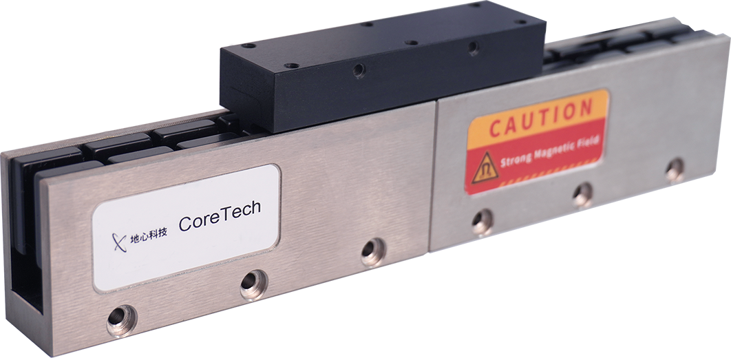
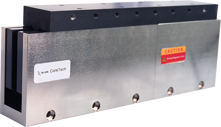
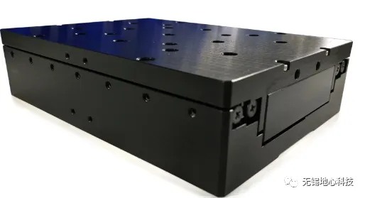
ART180 Series Linear Stage
As a scanning axis, high-density position-triggered camera capture at 5 nm intervals ensures high-resolution, distortion-free images.
Cross-roller bearings guarantee extremely high speed stability for the scanning axis.
Travel >200 mm ensures completeness and continuity of image acquisition.
Flexible travel configurations accommodate inspection of samples of varying sizes, from mouse brain to monkey brain to human brain.
As the lower axis, it offers strong load capacity and shear stiffness.
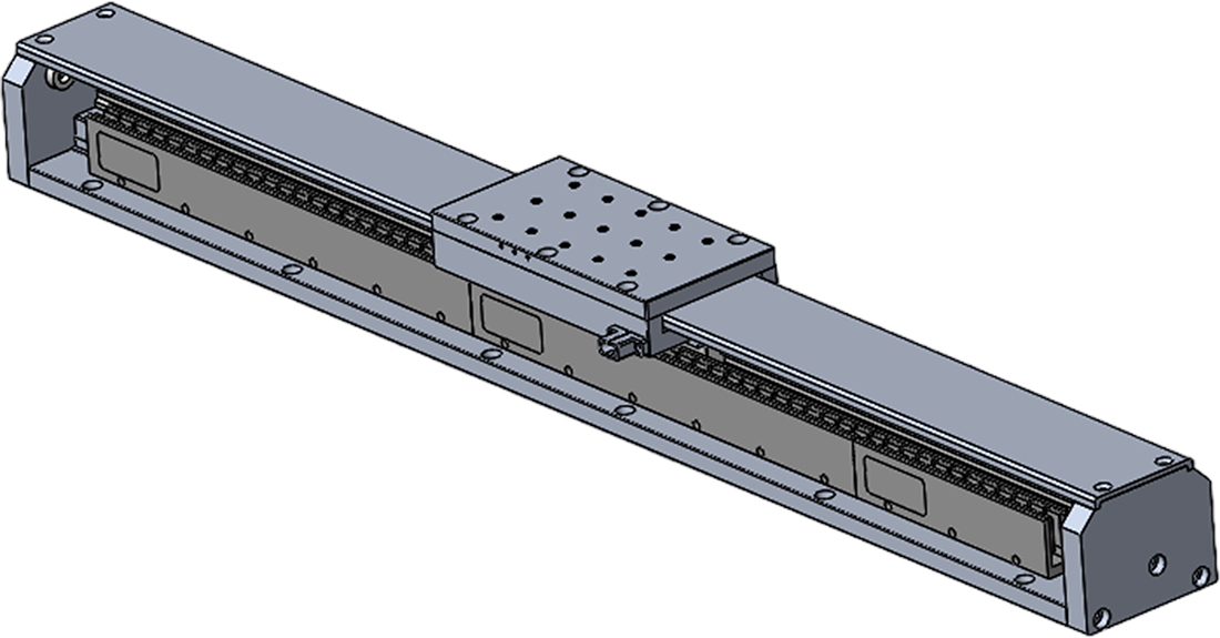
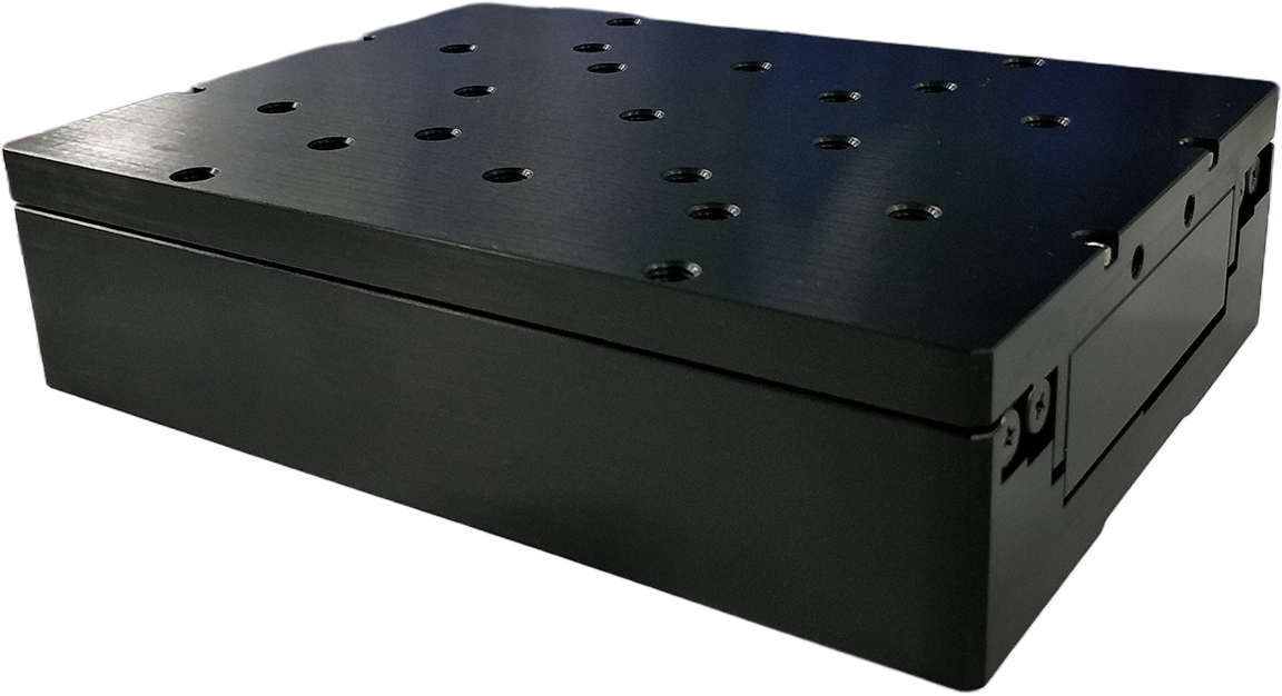
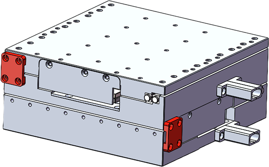
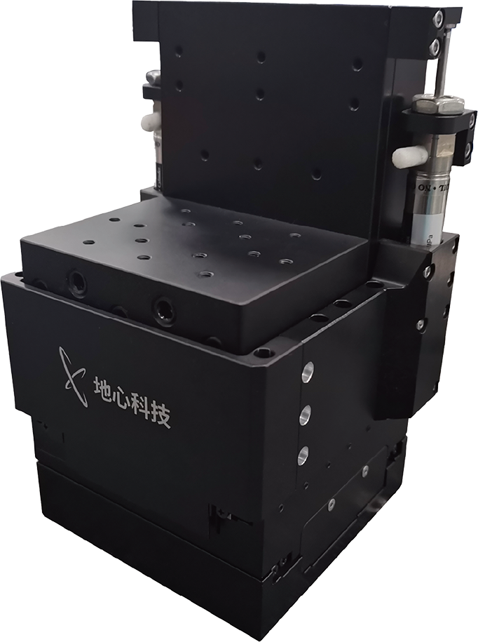
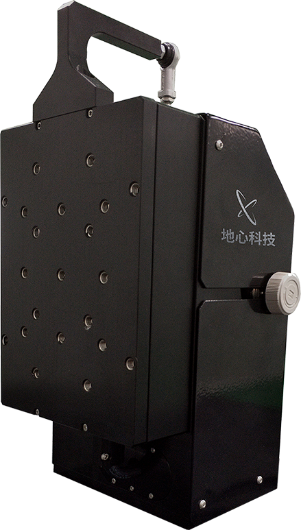
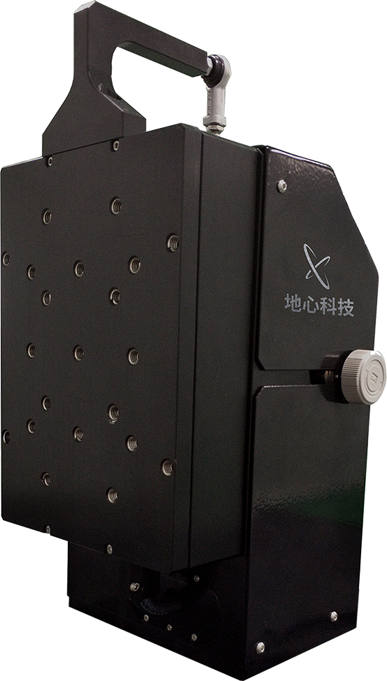
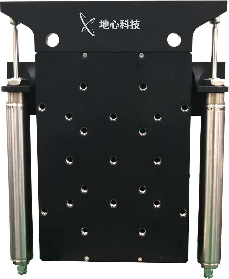
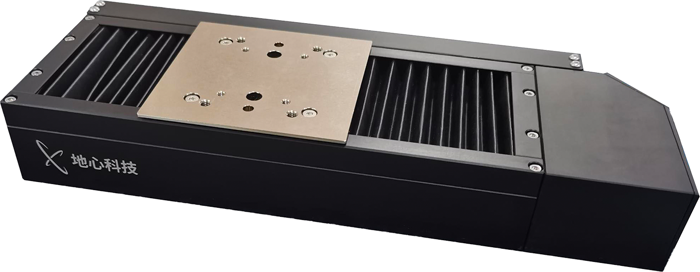
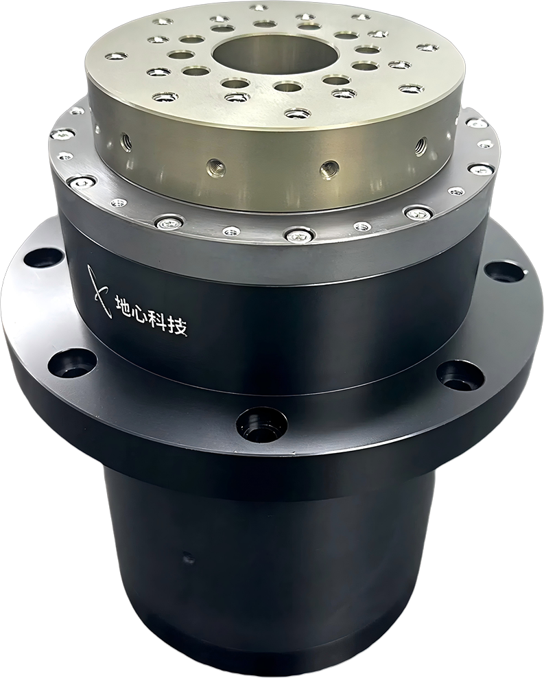
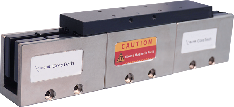
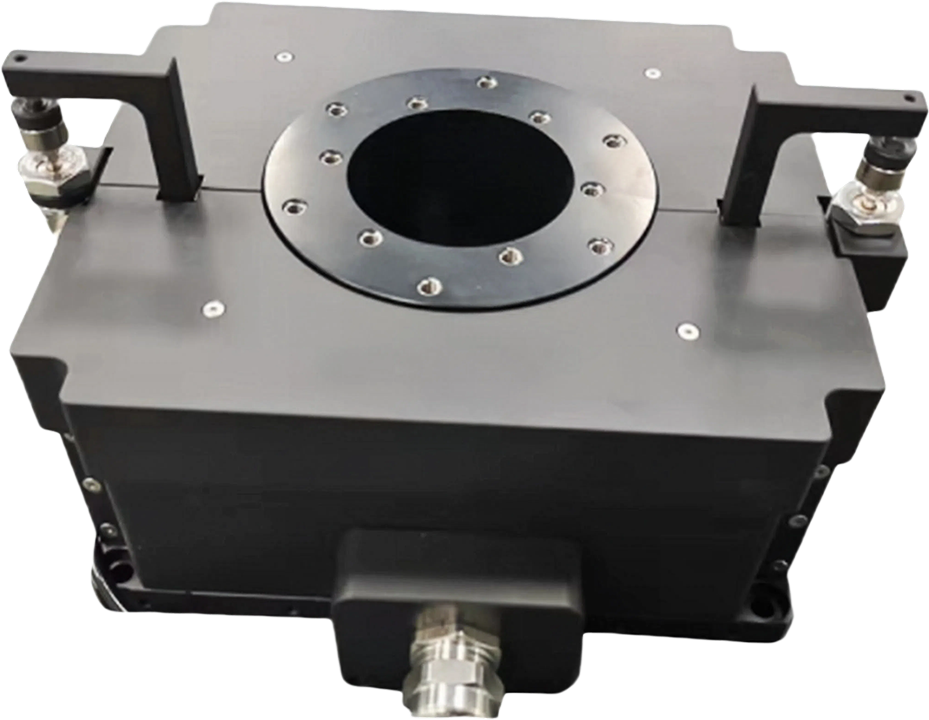
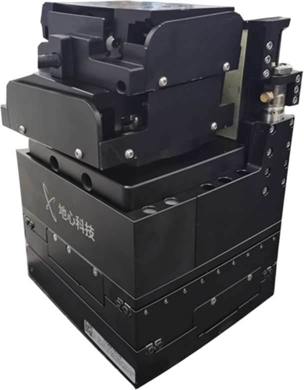
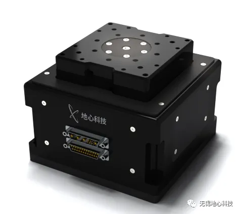
SMH165V Series Z-Lift Stage
As the lifting axis, minimum step can be less than 20 nm, ensuring small sectioning intervals.
Cross-roller bearings ensure high load capacity and excellent Z-axis straightness.
Robust structure withstands significant lateral blade cutting forces.
Optional travel configurations from 20 mm to 95 mm accommodate inspection of samples of varying sizes, from mouse brain to monkey brain to human brain.
04 Test Results
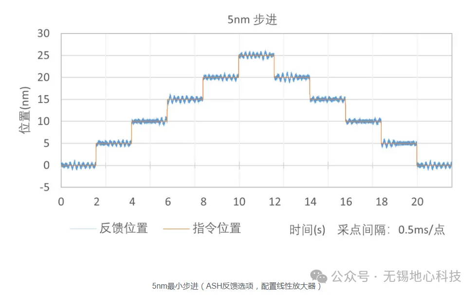
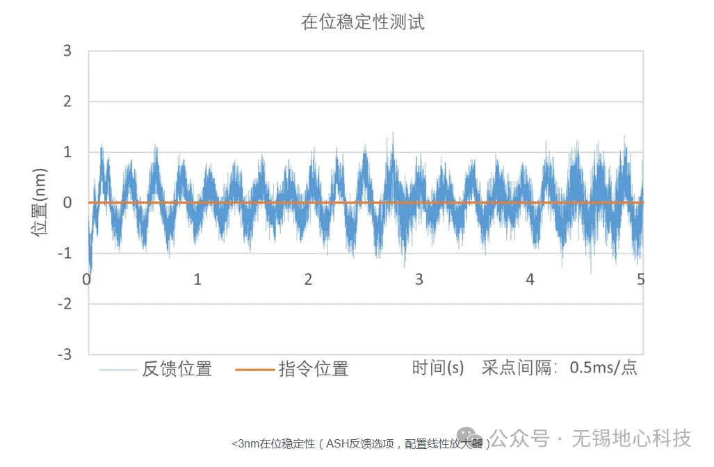
Taking the ART130 series as an example, despite its compact structure, the ART130 series stage offers strong driving capability, with maximum speed reaching 500 mm/s and no-load acceleration up to 1 g. Configured with a linear amplifier in a vibration-isolated laboratory environment, the ART130 series stage achieves in-position stability of 3 nm and a minimum step of less than 5 nm. Flexible customisation options are available based on customer application requirements.
Featured News
Beyond Cost and Energy Advantages: How Precision Motion Stages Further Enhance Nanoimprint Lithography Accuracy
2026.06.02
Semiconductor Wafer Inspection & IC Packaging Solutions – Sensor Chip Testing
2026.06.03
From Optical Chip Manufacturing to Fiber Coupling: Ultra‑Precision Motion Control Platforms Enabling a Technological Leap in the Optical Communications Industry
2026.06.03
 Back to List